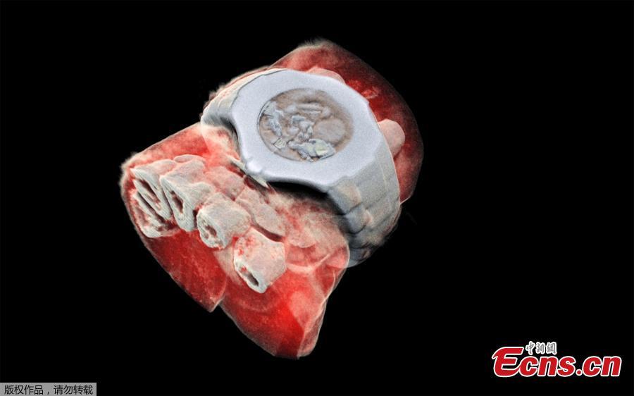
This picture released on July 12, 2018 by MARS Bioimaging Ltd shows a 3D image of a wrist with a watch showing part of the finger bones in white and soft tissue in red. New Zealand scientists has done the first-ever 3-D, colour X-ray on a human, using a technique promising to improve the field of medical diagnostics, announced Europe's CERN physics lab which contributed technology. (Photo/Agencies)
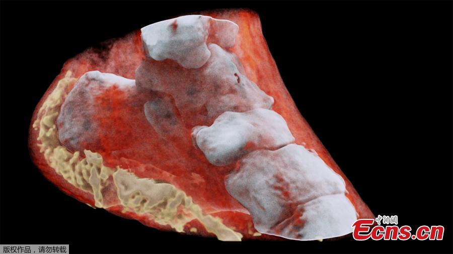
This picture released on July 12, 2018 by MARS Bioimaging Ltd shows a 3D image of left view of an ankle with bones in white and soft tissue in red. New Zealand scientists has done the first-ever 3-D, colour X-ray on a human, using a technique promising to improve the field of medical diagnostics, announced Europe's CERN physics lab which contributed technology. (Photo/Agencies)